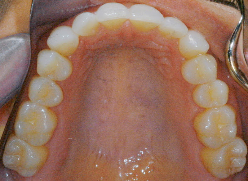
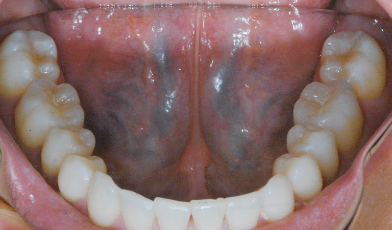
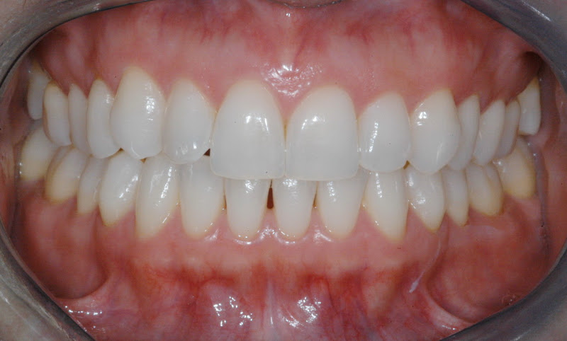
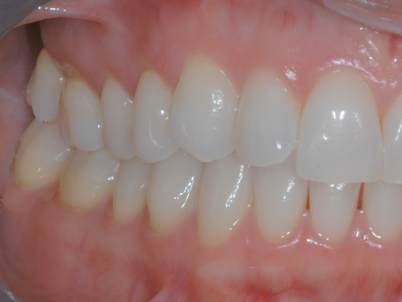
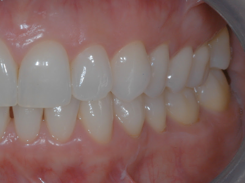
This patient is in her 30’s and has a dominant CR to CO discrepancy on the left side. Again in the anterior teeth frontal picture, note the position of the lower canines as compared to the uppers. The lower arch favors a class III bone relationship, but the mandibular teeth are tucked inside the upper teeth. The T-Scan Habitual Force Pattern is taken in the sitting or vertical head position. Another scan named the Skeletal Force Pattern is taken in the horizontal head position.
FUNCTION HEALTH
The Skeletal Force Scan is taken with the patient’s shoulders stretched back and the head supported either by a towel under the neck or the headrest. The skeletal scan gives critical information about the position of the bones without having to manipulate the condyle into centric relation. The difference between the vertical and horizontal scans is similar to your CR-CO slide.